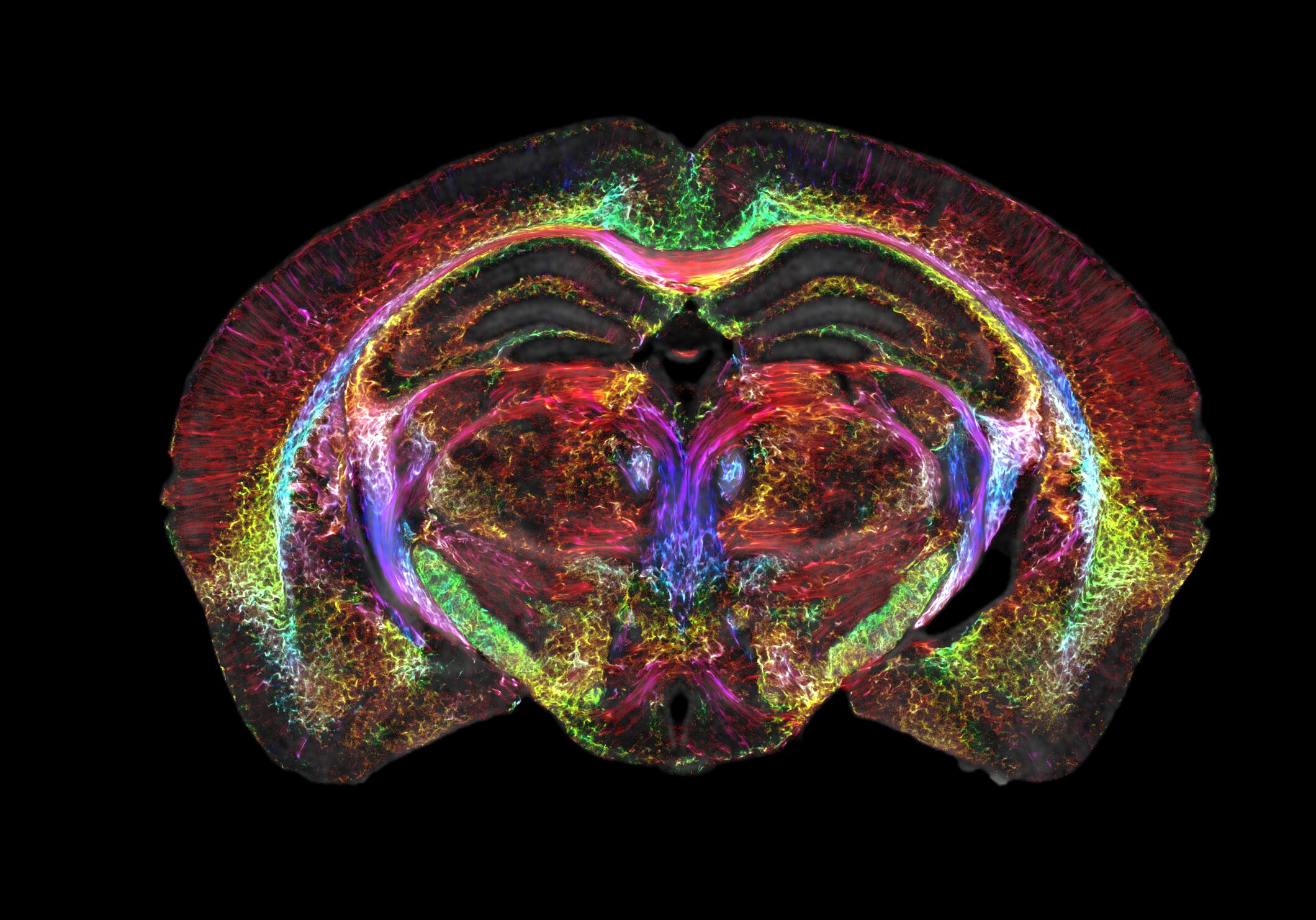
No, it’s not the latest psychotropic drug! Brain images just got 64 million times sharper.
Magnetic resonance imaging (MRI) is how we visualize soft, watery tissue that is hard to image with X-rays. But while an MRI provides good enough resolution to spot a brain tumor, it needs to be a lot sharper to visualize microscopic details within the brain that reveal its organization.
In a decades-long technical tour de force lead by Duke’s Center for In Vivo Microscopy with colleagues at the University of Tennessee Health Science Center, University of Pennsylvania, University of Pittsburgh and Indiana University, researchers took up the gauntlet and improved the resolution of MRI leading to the sharpest images ever captured of a mouse brain.
Coinciding with the 50th anniversary of the first MRI, the researchers generated scans of a mouse brain that are dramatically crisper than a typical clinical MRI for humans, the scientific equivalent of going from a pixelated 8-bit graphic to the hyper-realistic detail of a Chuck Close painting.
A single voxel of the new images—think of it as a cubic pixel—measures just 5 microns. That’s 64 million times smaller than a clinical MRI voxel.
Although the researchers focused their magnets on mice instead of humans, the refined MRI provides an important new way to visualize the connectivity of the entire brain at record-breaking resolution. The researchers say new insights from mouse imaging will in turn lead to a better understanding of conditions in humans, such as how the brain changes with age, diet, or even with neurodegenerative diseases like Alzheimer’s.
It is something that is truly enabling. We can start looking at neurodegenerative diseases in an entirely different way,” said G. Allan Johnson, Ph.D., the lead author of the new paper and the Charles E. Putman University Distinguished professor of radiology, physics and biomedical engineering at Duke.
Over the four decades, Johnson, his engineering graduate students and his many collaborators at Duke and afar refined many elements that, when all combined, made the revolutionary MRI resolution possible.
Some of the key ingredients include an incredibly powerful magnet (most clinical MRIs rely on a 1.5 to 3 Tesla magnet; Johnson’s team uses a 9.4 Tesla magnet), a special set of gradient coils that are 100 times stronger than those in a clinical MRI and help generate the brain image, and a high-performance computer equivalent to nearly 800 laptops all cranking away to image one brain.
full article: https://medicalxpress.com/news/2023-04-brain-images-million-sharper.html


Leave a comment